Abstract
Aesthetic and functional problems associated with significant facial asymmetry can negatively affect the patient's facial appearance, nutritional and psychosocial development. Therefore, a critical assessment and accurate treatment planning is absolutely necessary. The aim of this study was to evaluate the parameters of PA cephalometric analysis defined by Ricketts and Grummons and establish statistically relevant correlations and their importance in diagnosing orthodontic patients with varying degrees facial asymmetries. The research included facial asymmetry Romanian patients from the Department of Orthodontics and Dento-Facial Orthopedics of UMF "Victor Babes", Timisoara. The PA cephalogram investigations that met the inclusion criteria were digitally analyzed. Dental and skeletal cephalometric parameters described by Ricketts, Grummons and Kappeyne Van De Coppello were collected through linear, angular and volumetric measurements. Statistically significant correlations between the degree of asymmetry and the dimension of the internal structures were observed.Our conclusion is that PAcephalograms are cost effective and useful investigations in identifying and evaluating skeletal and dental imbalances in orthodontic facial asymmetry patients.
Author Contributions
Academic Editor: Sasho Stoleski, Institute of Occupational Health of R. Macedonia, WHO CC and Ga2len CC, Macedonia.
Checked for plagiarism: Yes
Review by: Single-blind
Copyright © 2020 Szuhanek C, et al.
 This is an open-access article distributed under the terms of the Creative Commons Attribution License, which permits unrestricted use, distribution, and reproduction in any medium, provided the original author and source are credited.
Competing interests
The authors have declared that no competing interests exist.
Citation:
Introduction
Facial symmetry is an important component of a person's attractiveness, representing one of the determining factors in its evaluation.1 Patients' concern for their physical appearance and for a more pleasing facial aesthetic is growing.2, 3 Thus, the orthodontist is more sought after than ever and often subjected to complex cases, which require a thorough examination to establish a correct diagnosis.4
The etiology of facial asymmetries is vast and in some cases remains mostly unknown.5 Morphology can differ under the prevailing environmental conditions under which the individuals’ development is taking place. 6, 7
Aesthetic and functional problems associated with significant facial asymmetry can negatively affect the patient's orofacial area, nutritional and psychosocial development. Therefore, a critical assessment and accurate treatment planning is absolutely necessary.8, 9, 10
Deviations from normal development, dento-maxillary anomalies in the transverse and vertical direction are studied in depth in the contemporary literature and, along with them, the correct evaluation of cephalometric measurements. 11, 12 As a complementary examination, the use of postero-anterior cephalometry was not introduced as early and used as widely as the lateral cephalogram.13, 14, 15, 16 but patients are three-dimensional, and thus the two dimensions visible on the lateral cephalogram should not be acceptable as the sole evaluations determining an orthodontic diagnosis. Numerous PA cephalometric analysis systems have been recommended each having strengths and limitations. Internationally, in countries such as Saudi Arabia and Peru, there is growing interest in the correct use and evaluation of postero-anterior cephalometric analysis parameters.17, 18 In 2011, Perez et. al used the Ricketts PA analysis to evaluate the norms of the Hispanic population.17 In 2019, the Grummons analysis was used in the symmetry assessment of the Saudi population in a study by Kumar et. al. 18
Aim and Objective
The main aim of this study was to evaluate and establish statistical correlations between a combination of PA cephalometric analyzes parameters defined by Ricketts and Grummons in a group of Romanian patients.
Material and Methods
This retrospective study included facial asymmetry Romanian patients aged 15 to 37 years, who presented at the Department of Orthodontics and Dento-Facial Orthopedics of UMF "Victor Babes", Timisoara between 2018-2020 and received orthodontic clinical examinations and complementary investigations such as: extraoral and intraoral photographs, study models, radiographs and cephalometric analysis. 50 patients had PA cephalograms made with the Cranex 3D device (Soredex). This research was conducted in the Orthodontic Research Center from Faculty of Dental Medicine, “Victor Babes” University of Medicine and Pharmacy, Timisoara.
The patient positioning system provided a head stabilization support, correct geometry and calibration ruler on each side of the image, teeth in maximum intercuspation position and a cephalometric light highlighting the Frankfurt plane that ensured the correct positioning of the patient. 35 PA investigations were excluded from the study due to not meeting the inclusion criteria: patients of Romanian origin (confirmed in the observation sheet) with no history of orthodontic treatment or trauma to the face, no craniofacial syndromes or skeletal deformities. The 15 remaining investigations were analyzed using the AudaxCeph software. Dental and skeletal cephalometric parameters described by Ricketts, Grummons and Kappeyne Van De Coppello were collected through linear, angular and volumetric measurements. The cephalometric reference points used in this study are presented in Table 1.
Table 1. Cephalometric reference points in Ricketts and Gummons PA cephalometric analisys| Cephalometric Reference Points | |
|---|---|
| ZL/ZR | The innermost point on the fronto-zygomatic suture |
| ZA/AZ | The outermost (lateral) point of the zygomaticarch |
| ANS | Anterior nasal spine |
| CN/NC | The outermost point of the nasal cavity |
| JL/JR | The highest point on the maxillary alveolar process |
| AG/GA | The deepest point of the antegonial depression |
| Me | The lowest point of the mandibular symphysis |
| A6/6A | The outermost point on the vestibular face of the upper molar |
| B6/6B | The outermost point on the vestibular face of the lower molar |
| B3/3B | The tip of the canine's cusp |
| CoR / CoL | The highest point on the mandibular condyle |
| Cg | Crista Galli |
| OccR / OccL | The point of first molar occlusion |
| A1 | The most marginal point at the incisal level of the upper central |
| B1 | The most marginal point at the incisal level of the lower central |
Dental parameters considered in this study are represented by molar relations on left and right (A6-B6 / 6B-6A linear measurement describing the vestibulo-lingual molar inclination), intermolar width and intercanine width. Skeletal relationships are defined by the following planes: right and left maxillomandibular width (JL / JR to ZL-AG / ZR-GA), maxilla-mandibular midline (angular measurement between the ANS-Me and ZA-AZ), maxillary width (JL-JR) and mandibular width (AG-GA). Dento-skeletal relationships were defined by the distance from the lower first molar to the JR-GA / JL-AG plane. The angle between the reference points ZA-AG-ZL / AZ-GA-ZR defined craniofacial relationship and the nasal width (NC-CN) described internal structures of the face. The reference points and planes of the Ricketts PA cephalometric analysis are illustrated in Figure 1.
Figure 1.Reference points and planes in the Ricketts analysis used to measure cephalometric parameters in facial asymmetry patients
To assess the discrepancies in facial asymmetry, three components of the PA analysis described by Grummons and Kappeyne Van De Kopello, which show left-right values, were generated by AudaxCeph version 6.0, after tracing the analysis, locating anthropometric points and tracing bone outlines: mandibular morphology - the analogous sides of the two mandibular triangles, formed between the points Co, Ag, Me and the two angles Ag (Co-Ag-Me) were compared; evaluation of linear asymmetries - the distance to the referencemidline and the difference between the vertical dimensions of the perpendicular projections of the bilateral landmarks (Co, NC, J, Ag) on the reference midline were calculated; maxillo-mandibular relationship - the distances from the vestibular cusps of the first maxillary molars to the projections of JL / JR to the reference midline were measured. In addition, the AG-AGplane, the ANS-Me and reference midline line were also drawn to reveal dental compensations for any skeletal asymmetry and the distances from the zygomatic-frontal sutures to the reference midline were measured and compared in order to evaluate the discrepancies in the upper floor of the face (Figure 2).
Figure 2.Transverse planes in the Grummons and Kappeyne Van De Kopello PA cephalometric analysis and evaluation of upper facial floor transverse asymmetry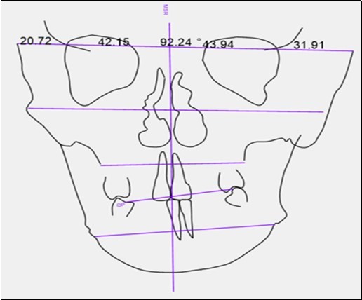
The MS Office Excel 16 was used to create the database and statistical analyzes were performed in the SPSS 24.0 program (SPSS, Chicago, IL). For the 15 selected measurements, the mean value and the standard deviation were calculated.
Results
Out of the total PA cephalometric analyzes, 40% men and 60% women, aged between 15 and 37 years. The mean age was 23 +/- 1 year for the women group and 21 +/- 1 year for men. The subjects were divided in groups based on gender and asymmetry severity.
After interpreting the measurements resulting from the Ricketts PA analysis, we observed that 30% of patients had a significantly deviated mandibular midline. The difference of the parameter values in the group of patients with significant asymmetries of the face compared to the other group, is represented in Figure 3.
Figure 3.Mean values for the mild asymmetry and the severe asymmetry groups
Comparison between patients with mild asymmetry and those with severe asymmetry, was done through the independent T test. The results found statistically significant differences in mandibular midline deviation, facial symmetry and nasal width (p <0.05).The independent T-test was also used to compare the differences between the sexes. There were no statistically significant differences between women and men.
To study the correlation in the Ricketts analyzes, the Pearson correlation coefficients were calculated. The coefficient with the highest value was between the mandibular width and the left maxillomandibular width (r = 0.929), and the one with the lowest value was found between the maxillary width and the left maxillomandibular width (r = 0.01). Significant correlations were found between intermolar and intercanine distance (r = 0.607) at p = 0.05. For the level p = 0.01 the significant coefficients with the highest value were found between the mandibular width (AG-AG) and the right maxillomandibular width (r = 0.644), and the significant coefficients with the lowest value turned out to be between the width maxillary (JL-JR) and and with the left maxillomandibular width (r = -0.752).
In the PA analysis described by Grummons, the following measurements on the two sides of the face were compared to the reference median-sagittal plane: mandibular morphology (Co-AG, AG-Me, Co-Me distances, AG angle); the asymmetry evaluation was performed by comparing the distances of the following points to the mid-sagittal line: Z, Co, Za, NC, J, AG and the left and right maxillomandibular relationship. The paired t test was calculated between the left and right measured values, but none of these were statistically significant. Pearson correlation coefficients were calculated to study the correlations. The coefficient with the highest value was between the AG angle and the Co-Me distance, both on the left (r = 0.639), and the coefficient with the lowest value at p = 0.05 was observed between the left Co-Me distance and the right Co-AG distance (r = 0.521). Other statistically significant values at the same p level were between the coefficients Co-AG right with Co-AG left, Co-Me left with Co-Me right, Co-Me right with Co-AG right and AG-Me right. At p = 0.01 the coefficients with the highest value were between the distance J right and the distance J left (r = 0.993), and those with the lowest value were observed between the angle AG on the left and Co-Me on the the right side (r = 0.651).
Discussion
Standardized methods of frontal cephalograms analysis have been around for decades most of them mainly used in surgical approach cases.13, 19, 20
Postero-anterior digital cephalometric analysis is accurate and especially useful in diagnosing asymmetry patients with transverse and vertical anomalies.21 The earlier these abnormalities in the development of the maxillary bones are discovered, the more beneficial it is for patients, allowing the initiation of a rebalancing treatment. This can avoid the need for dental camouflage treatments or more complicated types of orthognathic surgery.22 If skeletal imbalances are detected in patients who are still growing, immediate initiation of orthopedic and functional treatment is recommended, as true correction could be possible during this period.23, 24 To enhance the amount of data collected and surpass the limitations of the individual analysis, our study combines parameters from orthodontically oriented PA analyzes: Ricketts, Grummons and Kappeyne Van De Coppello.
To date, research in the field of antero-posterios cephalogram analysis focuses mainly on verifying the applicability of certain reference values in different populations.17, 18 Few studies focus on the correlations between various parameters. 25, 26 Patients included in this study that met the inclusion criteria exhibited varying degrees of facial asymmetry. When correlating gender with the evaluated parameters in our study no differences between the sexes were statistically significant in either of the PA analyzes. Our literature search found studies that found female faces to be more asymmetrical while others found no statistical differences between genders, which is in accordance to our findings. 27, 28 Soft tissues that cover the skeletal components tend to minimize skeletal asymmetry, in our study gender differences in the asymmetry of skeletal and soft tissue components were not statistically significant. The Ricketts analysis revealed statistically significant differences between parameters that describe the degree of asymmetry, respectively the deviation of the maxillo-mandibular midline together with one of the parameters of the internal structures, namely that of the nasal width. The quantitative analysis of Grummons found a constant dominance of asymmetrical development on the right side in most of the compared parameters, meaning that all skeletal and facial structures were situated significantly higher on the right side than on the left. The lateral area of the jaw exhibited a higher degree of asymmetry than other components of the face in all subjects. The postero-anterior analysis described by Grummons and Kappeyne Van de Koppello proved reliable in the determination of the severity of the asymmetry, which is an influential factor in deciding orthognathic intervention and diagnosing a possible underlying TMD.29
Conclusion
Considering the results obtained from our study group, we conclude that our combination of postero-anterior cephalogram analyses provided data that showed some positive and statistically relevant correlations between the evaluated parameters in asymmetry patients useful in correct diagnosis and orthodontic treatment planning.
Acknowledgment
This research was conducted in the Orthodontic Research Center from Faculty of Dental Medicine, “Victor Babes” University of Medicine and Pharmacy, Timisoara.
References
- 1.Little A C, Jones B C, DeBruine L M. (2011) Facial attractiveness: evolutionary based research. PhilosTrans RSocLondBBiolSci. 366(1571), 1638-1659.
- 2.Imani M M, Jalali A, Ezzati E, Heirani Z, Dinmohammadi M. (2018) A decision-making process to undergo orthodontic treatment: a qualitative study. Patient preference and adherence,2:. 2243-2251.
- 3.E De Baets, Lambrechts H, Lemiere J, Diya L, Willems G. (2012) Impact of self-esteem on the relationship between orthodontic treatment need and oral health-related quality of life in 11 to 16-year-old children. , Eur JOrthod 34(6), 731-737.
- 4.Borzabadi-Farahani A. (2012) A review of the evidence supporting the aesthetic orthodontic treatment need indices. , ProgOrthod 13(3), 304-313.
- 5.Smith R J, Bailit H L. (1979) Prevalence and etiology of asymmetries in occlusion. , AngleOrthod 49(3), 199-204.
- 7.Perrett D I, Burt D M, Penton-Voak I S, Lee K J, Rowland D A et al. (1999) Symmetry and human facial attractiveness. , Evol. Hum.Behav 20, 295-307.
- 8.Agrawal M, Agrawal J A, Nanjannawar L, Fulari S, Kagi V. (2015) Dentofacial Asymmetries: Challenging Diagnosis and Treatment Planning. , J Int Oral Health 7(7), 128-131.
- 9.Ko E W, Huang C S, Chen Y R. (2009) Characteristics and corrective outcome of face asymmetry by orthognathic surgery. , J OralMaxillofacSurg 67(10), 2201-9.
- 10.Dong Y, Wang X M, Wang M Q, Widmalm S E. (2008) Asymmetric muscle function in patients with developmental mandibular asymmetry.Journal of Oral Rehabilitation;35(1):. 27-36.
- 11.Mishra H, Shivaprakash G, Maurya R K. (2014) Assessment of Facial Asymmetry in Various Malocclusion: A Comparative Analysis. , J 48(4), 537-545.
- 12.De RosaA PerilloL, IaselliF. (2014) Comparison between rapid and mixed maxillary expansion through an assessment of dento-skeletal effects on posteroanterior cephalometry. Prog Orthod. 15(1), 46.
- 13.Broadbent B H. (1931) A new X-ray technique and its application to orthodontia.The Angle Orthodontist.1(2):. 45-66.
- 14.Steiner C C. (1960) The use of cephalometrics as an aid to planning and assessing orthodontic treatment: report of a case.American. , Journal of Orthodontics 46(10), 721-35.
- 15.Downs W B. (1952) The role of cephalometrics in orthodontic case analysis and diagnosis.American Journalof Orthodontics. 38(3), 162-82.
- 17.Kumar H, Kundi I, Alam M. (2019) Asymmetry Assessment for Saudi Adult Male Population: Grummons Analysis.World. , Journal of Dentistry; 10(1), 46-51.
- 18.Pérez I, Chávez A, Ponce D. (2011) Cephalometric norms from posteroanterior Ricketts' cephalograms from Hispanic Americans Peruvian non adult patients.Acta Odontol Latinoam. 24(3), 265-71.
- 19.Zwemer T J. (1976) Lorber RM: An annotated atlas of facial analysis,Dent. , Clin. N. Am 20, 641-660.
- 20.Grummons D C, MA Kappeyne van de Coppello. (1987) A frontal asymmetry analysis,J ClinOrthod;. 21(7), 448-65.
- 21.Sangroula P, Sardana H K, Kharbanda O P, Duggal R. (2018) Comparison of reliability and validity of posteroanterior cephalometric measurements obtained from Auto CEPH© and Dolphin® cephalometric software programs with manual tracing. , J Indian Orthod Soc; 52, 106-14.
- 22.Nagib R, Szuhanek C. (2020) Dentoalveolar compensation assessment using PA cephalon grams in orthodontic asymmetry patients. , Medicine in Evolution 1, 158-164.
- 23.Rubenduz M, Uslu O. (2010) Functional treatment of an asymmetry case having left side paralysis: a case report. , Eur J Dent 4(3), 341-347.
- 24.Srivastava D, Singh H, Mishra S. (2018) Facial asymmetry revisited: Part II-Conceptualizing the management. , Journal of Oral Biology and Craniofacial Research; 8(1), 15-19.
- 25.Athanasiou A E, Droschl H, Bosch C. (1992) Data and patterns of transverse dentofacial structure of 6- to 15-year-old children: A posteroanterior cephalometric study. , Am J Orthod Dentofacial Orthop 101(5), 465-71.
- 26.Snodell S F, Nanda R S, Currier G F. (1993) A longitudinal cephalometric study of transverse and vertical craniofacial growth. , Am J Orthod Dentofacial Orthop 104(5), 471-83.
- 27.Kaipainen A, SieberK. (2016) Regional facial asymmetries and attractiveness of the face. , European Journal of 38(6), 602-608.
Cited by (1)
- 1.Ganesan Poornima, Golla Usha Rao, Balashanmugam Baskaranarayanan, Munuswamy Geetha Lakshmi, 2024, Evaluation of Malocclusion Types in Adult Patients with Nasal Septal Defects – An Observational Cross-Sectional Analysis, Journal of Pharmacy and Bioallied Sciences, 16(Suppl 2), S1147, 10.4103/jpbs.jpbs_408_23